
Transforming Breast Health using Next Generation AI.
Patients are more informed than ever and are actively seeking breast health care providers who use cutting edge technology to help improve mammography quality and breast cancer screening effectiveness.
As patient volumes surge and health facilities face a critical shortage of radiologists and technologists, imaging centers often face the growing challenge of managing increasing workloads without compromising quality. Leading mammography facilities and clinicians leverage AI to improve workflows and limit the amount of rescanning and reading necessary for patients, and detect breast cancer faster, earlier, and more accurately, regardless of breast density.
Not all mammograms are created equal.
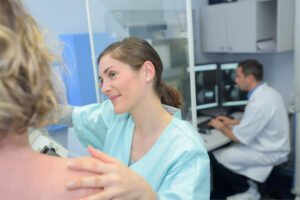 When a woman goes in for her screening mammogram, she assumes that the technologist can take a high quality scan for the radiologist to review. However, taking a scan is often a technically challenging task where positioning errors can often occur. Improving clinical image quality by using AI to assist technologists in evaluating positioning criteria provides higher quality images for radiologists and AI detection support solutions to more effectively detect breast cancer. The result, fewer unnecessary patient recalls due to unmet positioning criteria and inadequate clinical image quality.
How does a high-quality image boost efficiency & compliance?
High-quality screening images lead to higher mammography sensitivity and specificity, reduced costs, MQSA compliance, and ultimately better patient outcomes.
91%
of clinical image failures due to poor positioning1
75%
time saved on EQUIP/ACR processes2
50%
of mammograms would fail accreditation reviews3
Quality First: Better Image Quality Means Better Diagnoses
IntelliMammo® AI Platform powered by Densitas® provides continuous quality improvement tools for real-time and retrospective feedback for radiological technologists through synchronous real-time feedback at the point of care and asynchronous retrospective feedback at scheduled reviews. This continuous feedback helps technologists improve their positioning and capture screening mammograms accurately, ensuring higher clinical image quality from the start.
This enhanced clinical image quality means that radiologists have greater diagnostic confidence in their exam reviews and that resources can be focused on patients who genuinely need further evaluation and treatment instead of false-positive cases.
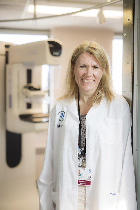
“We’re only as good as the images that we’re provided. So if we have images that are poor quality, we don’t know if we have missed any cancer on mammograms. So it’s critical that we have good quality images. I cannot stress that enough. And that’s why intelliMammo® is so useful for us as radiologists.”
Jean Seely, MD
Head of Breast Imaging, The Ottawa Hospital
Operational Efficiency: Continuous Improvement and Reduced Costs
IntelliMammo® AI Platform also offers a comprehensive mammography positioning technique knowledgebase with expert-prepared content for technologists. This enables continuous learning and development within the organization. Advanced analytics allow facilities to identify areas for improvement at the technologist and site level so that lower-performing technologists can be paired with higher-performing staff for timely and cost-effective training. Evidence-based standardized performance indicators support unbiased, efficient, regular, and cost-effective communication with technologists.
MQSA/ACR Compliance: Automate Tedious and Labor-Intensive Preparation for Audits
IntelliMammo® AI Platform automates mammography quality report generation, freeing up healthcare professionals to focus more on patient care rather than sifting through countless images to identify ACR quality studies. Advanced analytics track and assess technologist performance, allowing facilities to easily flag inadequate mammograms and trigger corrective actions that can be tracked to completion.
How can AI help detect breast cancer earlier and save lives?
 Detecting breast cancer early is vital; the earlier it is found while it’s still localized, the more treatment options exist, drastically improving patient outcomes.
ProFound AI® Breast Health Suite powered by iCAD helps radiologists concurrently read the high-quality screening images to help quickly focus their attention on regions of potential concern. It compares the findings across multiple slices and views, and it identifies a lesion score for each area it wants the radiologist to further evaluate.
How does the AI identify an area of concern?
The ProFound AI algorithm has undergone extensive training using one of the most comprehensive 3D image datasets available. Developed from 6+ million training images, ~8,000 biopsy-proven cancers, and 100+ contributing centers ensuring data diversity (as of June 2024), the AI algorithm provides radiologists proven data recommendations to inform their treatment plans.
The Art of Medicine & the next generation Science of Radiology.
Radiologists are trained experts in reading mammograms, and AI is their trusted partner to improve their reading specificity and sensitivity and the overall patient experience. By combining the detailed analysis from ProFound AI with the expertise of radiologists, the accuracy of cancer detection is significantly enhanced.5,6 This synergy improves patient outcomes, reducing the chances of missed diagnoses, and ensures timely treatment for those with breast cancer.
ProFound AI evaluates the lesion scores and breast density and provides an overall case score indicating the level of suspicion of breast cancer based on the screening population. These scores support radiologists in making more accurate treatment plans, balancing AI-driven analysis with their clinical expertise.
“ProFound AI offers a triple win: it helps radiologists detect more cancers, with fewer unnecessary callbacks, and in less time. This technology is poised to transform breast cancer detection and improve patient lives in the years ahead. I would not send a loved one to a mammography center that didn’t have ProFound AI, and there is no way, having experienced the functionality of the software, that I would ever read another tomo study without it.”
Mark Traill, MD
University of Michigan Health-West

Elevating Patient Care through Quality Images & Precise Detection.
IntelliMammo® AI Platform and ProFound AI® Breast Health Suite work together seamlessly with the imaging center’s existing equipment to provide a comprehensive solution to enhance mammography quality. In tandem they improve workflow efficiency, image quality, and diagnostic accuracy, leading to better patient outcomes and more efficient use of resources for cost-effective and sustainable breast screening service delivery.
The combined effect of better image quality and better detection leads to elevated patient care by reducing false positives and false negatives and increasing the sensitivity and specificity of mammography. This results in fewer unnecessary follow-ups, a decrease in unnecessary radiation exposure, and fewer invasive procedures.
In turn, patient satisfaction is higher, and patients have an improved experience and increased confidence in breast screening programs, leading to higher breast screening participation rates which is crucial for early detection and treatment, ultimately saving more lives.
Ready to learn more?
Learn more about Densitas and iCAD solutions and discuss how we can help improve the lives of your clinicians and patients.
To learn more about how intelliMammo® AI Platform and ProFound AI® Breast Health Suite can help improve image quality and accuracy of cancer detection at your facility, let’s connect.
References:
1. https://www.fda.gov/radiation-emitting-products/mqsa-insights/poor-positioning-responsible-most-clinical-image-deficiencies-failures
2. Densitas customer profile: Mammography facility processing 50,000 mammograms annually.
3. Guertin MH, et al. Clinical image quality in daily practice of breast cancer mammography screening. Can Assoc Radiol J. 2014 Aug;65(3):199-206. doi: 10.1016/j.carj.2014.02.001. Epub 2014 Jun 16. PMID: 24947189
4. https://www.nationalbreastcancer.org/breast-cancer-facts
5. Conant EF, Toledano AY, Periaswamy S, Fotin SV, Go J, Boatsman JE, Hoffmeister JW. Improving Accuracy and Efficiency with Concurrent Use of Artificial Intelligence for Digital Breast Tomosynthesis. Radiol Artif Intell. 2019 Jul 31;1(4):e180096. doi: 10.1148/ryai.2019180096. PMID: 32076660; PMCID: PMC6677281.
6. Schilling, K. Real-World breast cancer screening performance with digital breast tomosynthesis before and after implementation of an artificial intelligence detection system. Research presentation presented at European Congress of Radiology (ECR) 2023; March 1-5, 2023; Vienna, Austria.