Elevating Accuracy with ProFound AI® at Baptist Health South Florida

Kathy Schilling, MD
Medical Director, Christine E. Lynn Women’s Health & Wellness Institute, Boca Raton Regional Hospital
Challenge:
Facility upgraded to digital breast tomosynthesis (DBT), and the increase in reading volume introduced workflow challenges for radiologists aiming to detect cancer at its earliest possible stage.
Solution:
ProFound AI for Digital Breast Tomosynthesis and ProFound AI Risk*
Results:
ProFound AI empowered nine dedicated breast radiologists to find 23% more cancers, without increasing the rate of recalls. ProFound AI Risk personalized assessments for patients and supports clinicians in tailoring screening recommendations for women.
Summary:
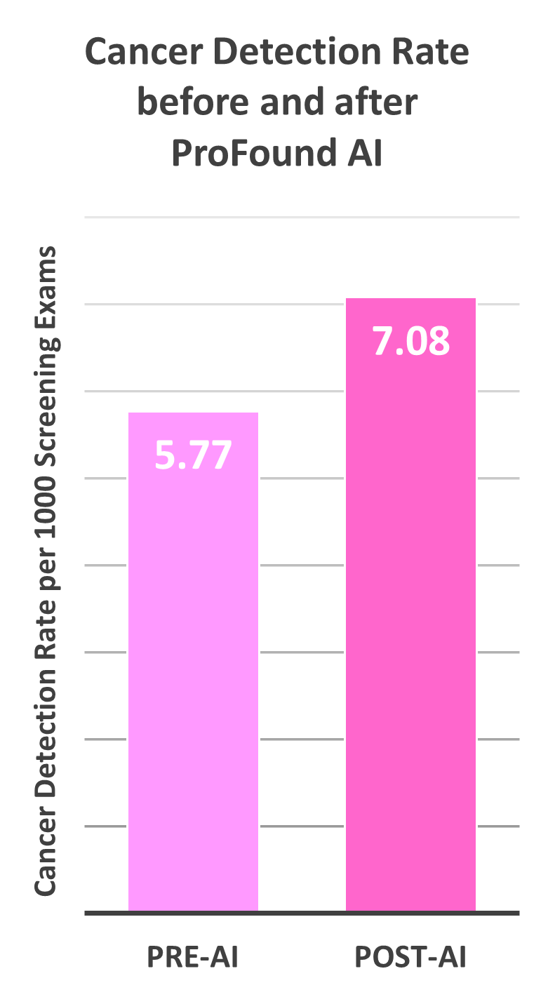
ProFound AI improved the cancer detection rate at the Christine E. Lynn Women’s Health & Wellness Institute by 23%, without increasing the rate of recalls.
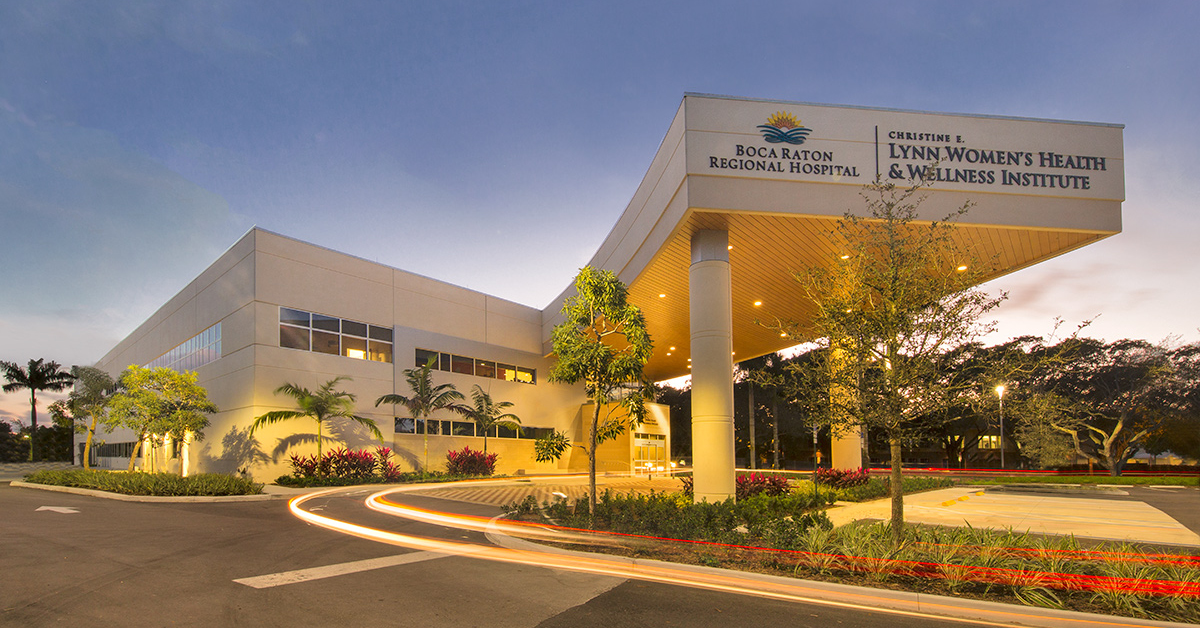
The story of the Christine E. Lynn Women’s Health & Wellness Institute, Boca Raton Regional Hospital
Part of the Baptist Health South Florida System, the Christine E. Lynn Women’s Health and Wellness Institute at Boca Raton Regional Hospital located in Boca Raton, FL offers a full range of women’s health care services, including breast cancer screening and diagnostic imaging. With three fixed sites and a mobile mammography van, the facility is equipped with 11 GE DBT Pristina units across its enterprise, and exclusively offers digital breast tomosynthesis (DBT) for both screening and diagnostic imaging.
Watch Webinar On-Demand
“We were really shocked at the outcomes of our research. With nine dedicated breast radiologists, all we do is read mammograms and breast imaging exams. With ProFound AI®, all our radiologists improved – across the board –
which was amazing to me.” – Kathy Schilling, MD
The Institute is led by board-certified breast imaging radiologist Kathy Schilling, MD, who has served as medical director at the facility since its inception in 1990. Under Dr. Schilling’s leadership, the facility has developed into the area’s premier and most preferred breast program, performing nearly 60,000 screening and diagnostic procedures a year. Dr. Schilling’s team consists of nine MQSA-certified, fellowship trained and dedicated breast imagers with an average of 22 years of experience.
This team performs more than 25,000 screening exams each year, providing results and personalized risk evaluations for women in real time.
“We are committed to providing our patients with the highest quality health care, which includes ensuring that our facility’s technology is the latest and we have the most experienced staff members on board,” said Dr. Schilling. “This included upgrading to DBT in 2014.”
When the facility upgraded to DBT, the team soon realized their work increased exponentially, as DBT yields hundreds of images per patient, compared to 2D mammography, which yields four images per patient.
“When we upgraded to DBT, we were suddenly reading tens of thousands of images a day, which can make it quite difficult to focus. It is mesmerizing to scan through a mammogram looking for a potential lesion that can be very small or subtle. Over time this can cause fatigue, which can ultimately result in burnout of our radiologists. That’s something we certainly cannot afford – we need every radiologist on our team working efficiently,” she added.
To overcome this challenge, Dr. Schilling advocated for the facility to adopt iCAD’s ProFound AI, which became the first AI technology for breast imaging to be FDA cleared in 2018. Built with the latest in deep-learning artificial intelligence, ProFound AI is clinically proven to improve radiologists’ sensitivity by 8%, reduce the rate of false positives and unnecessary callbacks by 7%, and slash reading time by more than half.1
“With all of our screening and diagnostic exams being DBT, we needed help managing the volumes of images we were seeing daily. We knew ProFound AI was clinically proven to help radiologists review cases more efficiently and with greater accuracy – and it certainly lived up to its promise,” said Dr. Schilling.
Trustworthy technology in uncertain times
The installation of ProFound AI was completed at the Institute in March 2020, and although the facility paused on screening for several weeks at the height of the pandemic, they were back up and running in May and immediately began to experience the power of ProFound AI.
“We were really excited to use the technology, and when breast cancer screening resumed at our facility in May, we jumped in right away and immediately confirmed its value,” said Dr. Schilling.
“Not only did ProFound AI improve our confidence reading DBT cases, but it improved our accuracy and efficiency. Even when we were seeing fewer patients at the height of the pandemic, ProFound AI helped our team find more cancers,” Dr. Schilling continued. “Additionally, when our patient volume returned to pre-pandemic levels a few months later, we were suddenly faced with a significant backlog of women who urgently needed a mammogram. ProFound AI helped us manage this influx in volume by empowering our team to read even complicated datasets faster and with more confidence.”
After using the technology for two years, Dr. Schilling was curious to learn more about the long-term impact of ProFound AI on her team, facility, and patients. She conducted a retrospective analysis of screening exams to compare the performance of her radiologists two years before and two years after adopting ProFound AI. She was surprised to discover ProFound AI helped her team find 1.31 more cancers per 1,000 screening exams for a 23% relative increase in cancer detection rate, without increasing the rate of recalls.2
Finding the cancers radiologists fear the most
Dr. Schilling not only confirmed ProFound AI was helping her team to find more cancers, they also noticed that the technology was finding the types of cancers radiologists fear the most.
“With ProFound AI, we are finding tiny lesions and subtle architectural distortions, which can be among the most difficult types of lesions to find. Many of these are invasive lobular carcinomas, which are notoriously difficult to spot and often present late,” said Dr. Schilling. “We’re starting to find cancers so early at our facility that, in many cases, women are able to avoid a mastectomy or chemotherapy. This is groundbreaking technology that is truly improving outcomes for our patients.”
Dr. Schilling’s team also adopted ProFound Risk, the first technology that provides an accurate, short-term risk evaluation based only on a mammogram.
“Breast cancer risk is typically evaluated based on factors such as age, breast density and family history, which are not modifiable. And, about 85% of breast cancers occur in women who have no family history of breast cancer.3 Our ability to detect cancer at the earliest possible stage begins with a precise understanding of a woman’s personal risk,” said Dr. Schilling.
“Recent studies confirm ProFound Risk is significantly more accurate than traditionally used risk models, both for long-term and short-term risk assessments.4,5 By offering personalized risk evaluations for our patients, our team is empowered to find cancers earlier, improve outcomes, and provide the best quality patient care,” Dr. Schilling continued.
References:
1. Conant, E et al. (2019). Improving Accuracy and Efficiency with Concurrent Use of Artificial Intelligence for Digital Breast Tomosynthesis. Radiology: Artificial Intelligence. 1 (4). Accessed via https://pubs.rsna.org/doi/10.1148/ryai.2019180096 2. Schilling K. Real-world breast cancer screening performance with digital breast tomosynthesis before and after implementation of an artificial intelligence detection system. Presentation at European Congress of Radiology (ECR) meeting; March 2023; Vienna, Austria. 3. U.S. Breast Cancer Statistics. Breastcancer.org. Accessed via https://www.breastcancer.org/symptoms/understand_bc/statistics. 4. Eriksson, M et al. A risk model for digital breast tomosynthesis to predict breast cancer and guide clinical care. Science Translational Medicine. 14 (644). 2022 May 11. Accessed via https://www.science.org/doi/10.1126/scitranslmed.abn3971 5. Eriksson M, CzeneK, Vachon C, Conant E, Hall P. Long-Term Performance of an Image-Based Short-Term Risk Model for Breast Cancer. Journal of Clinical Oncology. DOI: 10.1200/JCO.22.01564.