
ProFound Breast Health Suite
Breast cancer has finally met its match.
Powered by the latest innovations in artificial intelligence (AI), the ProFound Suite offers clinically proven, 360-degree solutions for cancer detection, density assessment, and personalized risk evaluation. ProFound offers unrivaled accuracy, as well as multi‐vendor compatibility and unique workflow advantages.

ProFound Breast Health Suite
Together, we can change more lives.
Backed by science, clinical evidence, and proven patient outcomes, our suite of solutions – Detection, Density, and Risk* – shine a spotlight on cancer, exposing its hiding place. Empowering you to personalize breast cancer screening like never before.
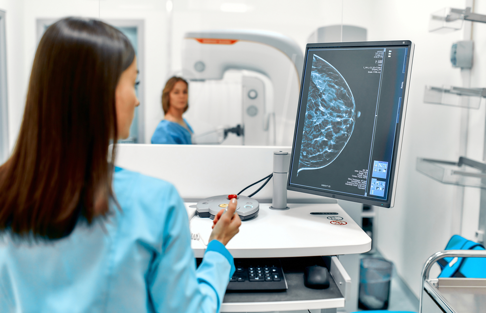
Cancer Detection
Built on one of the largest, most diverse global data sets, our Detection solution offers unrivaled performance and accuracy for both 2D and 3D mammography.4 Scoring cases and lesions so you can identify and focus on areas of most concern.
Density
The Density Assessment solution simplifies and standardizes breast density reporting and stratification, with accurate and reliable results. By analyzing 2D or 3D mammogram images, it examines the woman’s breast anatomy, delivering clinicians unique and patient-specific breast density assessments, so they can proactively implement a personalized screening plan.

Risk
Our Risk solution* is different, offering up to 2.4x more accuracy than traditional risk models like Tyrer-Cuzick and Gail,1,2 and is personalized based on age and image evidence directly from their mammogram. It’s near-term providing a risk probability score for developing breast cancer in the next 1 or 2 years giving women and care providers more actionable information to design personalized care plans.
ProFound AI Risk is CE Marked and Health Canada Licensed, is not FDA Cleared, and is only available in the US for investigational use.
Research Candidate
Breast Arterial Calcifications
Breast cancer and heart disease are the two leading causes of death among women. Clinical results have found calcifications in arterial vessels within the breast are proven to correlate with calcifications elsewhere in the body, which raises concern for cardiovascular health issues.
With our BAC Assessment solution, the presence and extent of breast arterial calcifications can be identified and measured.
Imagine a future where a single mammogram informs insights beyond breast health!

Expert Voices
Radiologists share their experiences using ProFound Breast Health Suite


Experience the ProFound AI difference.
As the clinically proven industry leader, radiologists using ProFound AI find more cancers more often, more accurately, with fewer unnecessary recalls.
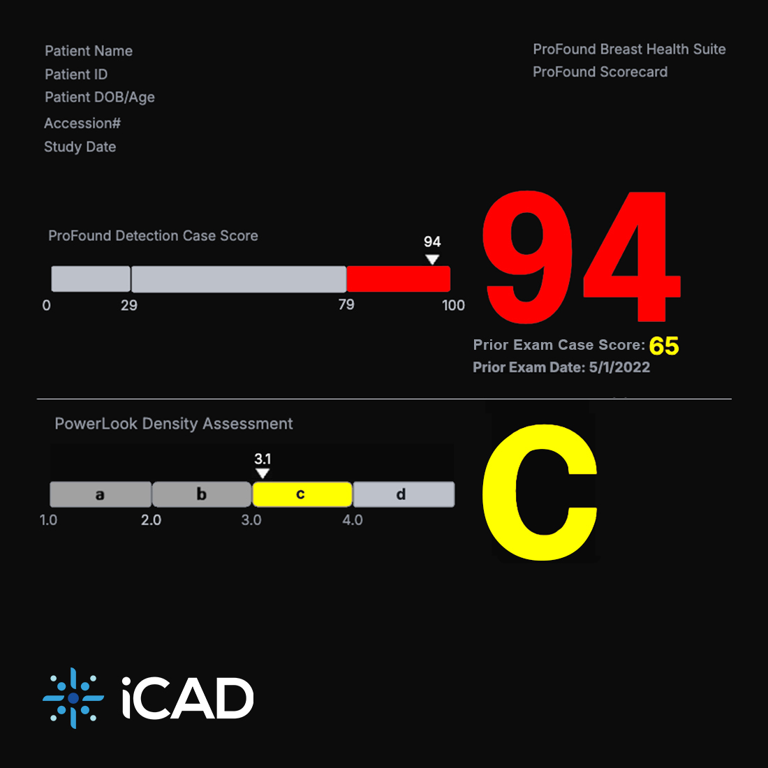
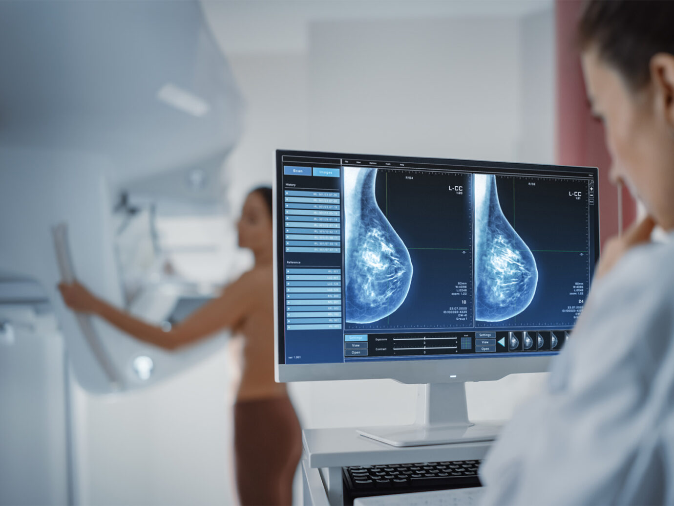
Example of ProFound Scorecard
Meet the ProFound Scorecard.
Automated, integrated, and easy to read – the ProFound Scorecard aids in early cancer detection, breast density assessment, and short-term risk evaluation via actionable mammographic case summary. Giving you the information needed to personalize patient care moving forward.
New features help breast imaging professionals quickly identify suspicious lesions by limiting the number of marks on each 3D view, and understand the level of suspicion related to lesions and the case through color-coded scoring ranges.
Includes: Overall Case Score. Automated Density Assessment Score. Personalized 1-2 year Risk Score.*
Worklist prioritization and color based on PACS viewer and output requirements.
ProFound AI Risk is CE Marked and Health Canada Licensed, is not FDA Cleared, and is only available in the US for investigational use.
Quality healthcare, reimagined.
Align with Quality Standards.
AI-powered mammography is playing a role in helping breast imaging centers align to important quality standards and accreditation programs.
- Mammography Quality Standards Act (MQSA)
- National Quality Measures for Breast Centers (NQMBC) Program
- National Accreditation Program for Breast Centers (NAPBC)
- Institute for Healthcare Improvement Quintuple Aim of Healthcare
- American College of Radiology (ACR) Accreditation Program.
Mark Traill, MD
My MQSA numbers are the best they’ve ever been. My productivity is as high as it has ever been, with this algorithm and with this suite of tools here. Everybody wants you to do a better job and work faster, and this has allowed me to do it.
Breast Radiologist, University of Michigan Health-West
ProFound Interoperability.
Flexible deployment with unmatched speed, scalability, and security.

ProFound Cloud
Your
Cloud

ProFound
On-Premises
50+ PACS & 94 versions | 2D & 3D OEM manufacturers | AI platform providers & workflow systems
We work the way you want to work.
Rapid implementation for a swift go-live.
Flexible & secure deployment options for smooth IT adoption.
Seamless integration within your workflow and systems you trust.


Contact Us
Together, we can create a world where cancer can’t hide.
Ready to experience how ProFound can save lives?
1. Mikael Eriksson et al. A risk model for digital breast tomosynthesis to predict breast cancer and guide clinical care. Sci. Transl. Med. 14, eabn3971 (2022). DOI: 10.1126/scitranslmed.abn3971. 2. Eriksson M, Czene K, Strand F, Zackrisson S, Lindholm P, Lång K, Förnvik D, Sartor H, Mavaddat N, Easton D, Hall P. Identification of Women at High Risk of Breast Cancer Who Need Supplemental Screening. Radiology. 2020 Nov;297(2):327-333. doi: 10.1148/radiol.2020201620. Epub 2020 Sep 8. PMID: 32897160. 3. Schilling K. et al. Real-world breast cancer screening performance with digital breast tomosynthesis before and after implementation of an artificial intelligence detection system. Presented at the European Congress of Radiology (ECR), March 1-5, 2023 in Vienna, Austria. 4. Conant EF, Toledano AY, Periaswamy S, Fotin SV, Go J, Boatsman JE, Hoffmeister JW. Improving Accuracy and Efficiency with Concurrent Use of Artificial Intelligence for Digital Breast Tomosynthesis. Radiol Artif Intell. 2019 Jul 31;1(4):e180096. doi: 10.1148/ryai.2019180096. PMID: 32076660; PMCID: PMC6677281. 17. Schilling et al. The Real-World Effect of Artificial Intelligence on Histopathology and Stage in Breast Cancer Screening with Digital Breast Tomosynthesis. Presented at the 2024 Radiological Society of North America (RSNA) Annual Meeting; December 1, 2024; Chicago, IL, USA.
iCAD data on file. Standalone performance varies by vendor.